Hip disorders in children and adolescents
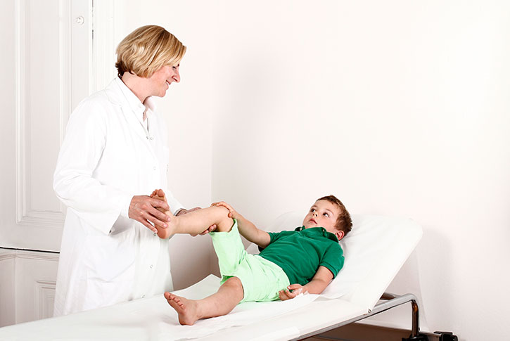
Early diagnosis is the key to successful treatment of any disorder. An exact clinical examination and radiographic tools like ultrasound, X-ray and magnetic resonance imaging help to establish the correct diagnosis. Typical signs of hip disorders in children are limping, flexion of the hip, groin pain, as well as pain projecting to the knee. Follow-up is as important as primary treatment. The development of the hip joint must be monitored until growth arrest in order to achieve optimal function in adulthood.
Hip dysplasia
- Poor development of the socket
- Disrupted ossification of the bony rim of the socket
- Insufficient coverage of the femoral head may lead to dislocation or instability of the hip Joint
Complaints: limited abduction of the hip joint, leg length discrepancy, limping, pain, early osteoarthritis
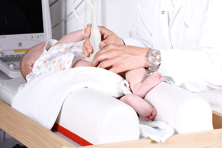
Prevention: Hip ultrasound screening and orthopedic examination in the 1st and the 6th to 8th week of the infant’s life (guidelines of the Austrian mother-child medical card)
Therapy: Abduction therapy with special bandages or splints are successful in some patients while others require more rigid fixation with a cast. Surgery is performed only in cases of severe dysplasia. Coverage of the femoral head is improved by corrective ostoeotomies in the femur and/or acetabulum. When hip dysplasia is identified too late or not treated on time it may be necessary to perform surgery in adulthood..
Coxitis fugax
Coxitis fugas is very common in toddlers and schoolchildren, and usually occurs after an infection of the respiratory, gastrointestinal, or urinary systems. It is a transient inflammatory reaction of the hip joint. The symptoms include limping, intolerance of weightbearing, as well as painful and limited motion.
Treatment: bed rest, antiinflammatory medication
Note: Septic arthritis of the hip joint caused by bacteria may look similar and should not be overlooked. In most cases it is accompanied by fever, swelling, redness, and very severe pain. Acute surgery with irrigation and drainage of the joint as well as antibiotic treatment are mandatory.
Perthes disease
Blood supply to the femoral head is compromised in Perthes disease, which is most common in children between the ages of 4 and 10 years. It occurs more frequently in boys than in girls. The femoral head loses its round shape, leading to deterioration of joint function and early degeneration of the joint.
Complaints: Intermittent symptoms in the thigh, such as limping, pain (which may also extend to the knee), limited range of motion, and leg length discrepancy.
Treatment: Physiotherapy and observation are sufficient in many cases. In patients with more severe deformation of the femoral head it may be necessary to perform surgery. The goal is to improve the alignment of the joint, which enhances the likelihood of good joint function and a rounded femoral head.
Slipped capital femoral epiphysis
The femoral head slips off the femoral neck in the region of the growth plate. This condition occurs most commonly in adolescents.
Complaints: The symptoms start slowly over a period of several weeks and may worsen suddenly. Pain, limping, outward rotation, and shortening of the leg may occur. The range of motion and weightbearing are limited.
Treatment: Surgery is essential. In mild slips the bone is fixed with a screw or wires to prevent further dislocation. In more severe slips with impaired joint function, the femoral head is repositioned and fixed with screws.
Congenital foot deformities
Congenital foot deformities may be encountered in infants. In most cases these are harmless malalignments that resolve on their own. Massaging the foot is a useful supportive measure. These deformities include talipes calcaneovalgus and metatarsus adductus.
In contrast, some congenital malformations call for special types of treatment.
Clubfoot
The clubfoot may occur on one side or both sides. Boys are affected more often than girls. The cause is unknown, hence the term “idiopathic clubfoot”. This is a malalignment of bone with simultaneous contraction of tendons and ligaments, which leads to inward rotation of the foot or a drop foot. Depending on its severity, this deformity may be partly expandable or fixed.
Treatment: The deformity is treated with plaster casts. The Ponseti method is a gentle type of treatment, in which the plaster cast extends from the foot to the thigh. The casts are changed once a week. After 6 to 8 weeks the deformity is corrected to the extent that just a contraction of calcaneal tendon (Achilles tendon) remains. This is treated by a minimally invasive operation.
In a so-called percutaneous tenotomy of Achilles tendon, a tiny skin incision is made to transect the tendon and thus extend it. This is followed by another plaster cast. Subsequent treatment with splints and braces is as important as correction of the deformity itself. After the last plaster cast, special braces are prepared to fix the feet in externally rotated position on a metal bracket. The braces have to be worn day and night (22 hours; they may be removed for body care) for 3 months, after which they need to be worn only at night. The brace treatment is continued until the age of four years. The treatment is accepted very well by the young patients as the braces do not hinder the child when crawling or learning to walk.
This method is very successful in most patients. Your child will be able to walk normally, play, and do sports. In cases of unilateral foot deformities, the calf may be somewhat thinner on the deformed side than on the opposite side and the shoe on this side may be one or two sizes smaller than the opposite shoe.
Sometimes the deformity may reappear after some time. In these cases the patients may be given renewed brace treatment or it may be necessary to relocate the tendon. Therefore, regular controls until the completion of growth are necessary.
A clubfoot may also occur in the course of various underlying diseases. These include neurological diseases or syndromes. One uses the Ponseti method in these special cases as well. However, as the foot deformities tend to be more severe in these cases, it may be necessary to perform more extensive surgery.
Pes planovalgus/flatfoot in children
A flattened longitudinal arch and outward deviation of the heel may cause the foot to give way or buckle when standing and walking. This type of foot is very common in infants and is a part of the normal development of the foot. In most cases the phenomenon resolves on its own during growth. Treatment is not necessary when the patient has no pain and his/her muscles are functioning well. This is tested by having the patient rise onto the toes. In cases of more pronounced deformities, which may also be accompanied by pain, the use of insoles and muscle strengthening is the first treatment step. Occasionally it may be necessary to perform minimally invasive surgery (Arthrorisis, calcaneus stop screw), which exerts a positive impact on the deformity while the foot is still growing.
The congenital flatfoot is a rare deformity which also needs to be treated. Depending on its severity one uses plaster casts or surgery for its correction.
Stiff flat feet are rarer and cannot be corrected adequately by muscle build-up or insoles. It is caused by underlying neurological diseases, disrupted ossification, or connective tissue disease; the cause must be included in the treatment plan.
Deformities of the extremities
Axis deviations (knock knees and bow legs), leg length discrepancies
The leg axis changes in the course of development. Slight bow legs are common during the first two years of a child’s life, knock knees are typical after the age of three years. Around the age of eight years the leg achieves its normal axis.
In cases of marked deviations from the normal condition for the respective age, various joints may be overloaded. This may be accompanied by disabilities in walking, and pain. In these cases the leg axis can be improved by surgery. In the growth phase it will be possible to regulate growth, so to speak, by temporary blockade of the growth plate (hemiepiphysiodesis). This is accomplished by the use of metal plates that curtail growth.
In patients with very severe deformities it may be necessary to perform surgical sectioning of bone (osteotomy) for the purpose of vertical adjustment, and then fixation with plates and screws. In cases of very severe deformities in several planes (kinking, length difference and twisting) one uses so-called external fixators. These are ring-shaped systems interconnected by telescopic rods and fixed in bone with metal pins. After exact planning of the procedure with the aid of a computer program, the patient is able to move the rings against each other by rotating the telescopic rods and thus achieve correction in several planes. Severe bone distortions, such as those after accidents, inflammation, tumors, or in cases if congenital skeletal disease, can be corrected by this method. The patient is able to achieve a normal standing position as well as the ability to walk normally.
Slight leg length differences of 1 cm or less are quite common and are of no consequence. Larger leg length differences must be investigated and monitored. Leg length differences have various causes. Different bone lengths may arise due to inflammation, injury, previous operations, tumors, or in a variety of skeletal diseases.
Length differences that led to a disruption of gait and deformities of joints or the spine must be treated. The treatment depends on the child’s age and the magnitude of the length difference. Insoles that balance the height difference or elevations of the shoe soles may compensate mild differences. In cases of larger length differences it will be necessary to perform surgery. Exact planning and the establishment of a growth prognosis are essential. Length differences can be treated by retarding the growth of the longer bone (epiphysiodesis) or elongating the shorter bone. In cases of so-called callus distraction the bone is transected. New bone (callus) is formed in the bone gap, which is then gradually lengthened. Once the desired length has been achieved the movements are stopped. The newly formed bone bridge is solid and capable of withstanding loads.
Bone lengthening is achieved by various techniques. External fixators or intramedullary nails introduced into bone and elongated in telescopic fashion are the methods of choice. During and after bone lengthening the patient must perform physiotherapy to preserve the mobility of all joints and ensure the functional capacity of tendons and ligaments.
Intoeing
Many children direct the forward section of the foot inward when walking. Such “intoeing” should not be regarded as an abnormality. Rather, it is caused by the development of the hip joint. During growth the femoral bone close to the hip joint gradually turns backward (detorsion). As a result the feet are rotated outward and the person’s gait is normalized. There are two spurts of growth: the first occurs around the age of 6 years and the second around the age of 11 years. During these periods the situation improves rapidly. Sometimes the rotational error is in the calf region. The differentiation is made by clinical investigation. Only in very severe cases – for instance when the child tends to trip over his/her own feet often or gets caught at the knees – it will be necessary to perform surgery.
The spine of the child and the adolescent
By performing orthopedic investigations after birth, at the preschool age and at the start of puberty, spinal curvatures and deformities can be identified on time. The clinical investigation will disclose changes. In most cases the changes call for further clarification, which is then achieved by obtaining X-rays or performing magnetic resonance imaging.
Scoliosis
Congenital changes occur in the presence of diseases of the muscles or the nervous system, as well as in metabolic diseases. However, so-called idiopathic scoliosis is the most common condition; it occurs primarily in girls and a familial clustering of the disease has been noted. The condition is marked by a distortion and malrotation of the spine, leading to asymmetry in the spine and the rib cage. It may even induce a height difference between the two shoulders and the iliac crests. Pain is rare. The treatment differs, depending on the patient’s age and the stage of development. In every case one must perform regular controls to detect any deterioration on time and initiate counter-measures.
Physiotherapy for strengthening the muscles of the trunk and the abdomen are useful in every stage. When the distortion exceeds a certain limit, corsets are used. This is successful as long as growth has not been concluded. Operations are necessary only in very severe forms of the diseases.
Spondylolisthesis
Back pain in a child or an adolescent may be caused by a so-called slipped vertebra or spondylolithesis. This condition is marked by greater mobility between two vertebral bodies, caused by congenital or developmental changes in vertebral bone. Gaps in the vertebral arch (spondylolysis) may be one cause. Pain occurs mainly in puberty and is typically more severe after heavy strain. Radiological investigations - X-rays and magnetic resonance imaging - help to determine the extent of spondylolisthesis and its potential effect on bone marrow or nerve roots. Treatment is required only when the patient experiences pain. First one should perform physiotherapy to build up the muscles. Sometimes temporary immobilization with a corset may alleviate the pain. In severe cases it will be necessary to perform surgery and achieve fusion of the slipped vertebra.
Disk prolapse
Disk prolapse is a rare event in children and adolescents. However, excessive strain or accidents as well as congenital changes in the spine may cause a prolapse of disk tissue (disk prolapse) even in this age group. This may lead to irritation of nerve roots or bone marrow. Pain radiating into the leg, loss of sensation or manifestations of paralysis are warning signs of this disease.
Knee joint, Cartilage damage
Knee pain in children and adolescents may have various causes. One typical cause is irritation of the tendon insertions, which mainly occurs during growth and sports. These include Osgood-Schlatter disease or Sinding-Larsen syndrome, which concern the ligaments and tendons in the region of the knee cap or patella. Another reason for knee pain may be disturbed blood circulation in the joint cartilage and in adjacent bone, which leads to a condition known as osteochondrosis dissecans. This condition is mainly observed in active boys. In many cases the disease heals spontaneously, but when followed by loosening of the bone and cartilage fragment it may have serious consequences on the joint, and may necessitate surgery. The treatment is selected according to the age and type of damage. In many cases it is enough to drill holes into the bone; in others in may be necessary to fix the loosened fragment with screws or use modern techniques like cartilage cell (chondrocyte) transplantation. Snapping phenomena or joint blockades may be caused by a discoid meniscus. The meniscus acts as a shock absorber of the knee and is usually C-shaped, but may be shaped like a disc in some persons. These variants are frequently associated with tears, which then cause complaints. The diagnosis is established by magnetic resonance imaging. The excess meniscus tissue is removed by performing an arthroscopy of the joint, and its normal shape is restored.
Inflammation, bone cysts, tumors
A purulent inflammation may occur in any part of the musculoskeletal system, i.e. muscles, bones, or joints. They are caused by bacteria which are then dispersed through the bloodstream. Fever, reddening, swelling and severe pain – even at night - are alarm signals. Quite often the children are generally weakened, exhausted, and tired. Rapid diagnosis with the use of imaging procedures and a blood investigation are decisive measures to initiate the appropriate treatment. In most cases one needs to perform early surgery and remove the center of the inflammation.
Bone cysts are cavities filled with fluid, which may be found in various bones. Quite often they occur close to joints. Sometimes they are discovered by accident or become evident due to a fracture, which may even occur without forceful impact because the bone is weakened by the cyst. The treatment differs according to the type of cyst. Small cysts can be monitored; in cases of large cysts one usually needs to perform surgery. In many cases one can use the minimally invasive approach.
Tumors of the musculoskeletal system are rare, but may be a threatening condition. It is important to distinguish between malignant and benign tumors. An X-ray and a magnetic resonance investigation do indicate the right condition, but the final distinction is made by obtaining a piece of tissue and performing an investigation of the tissue (histology). Specialized centers like the University Clinic of Orthopedics at the General Hospital of Vienna are the right place to establish the diagnosis and treat children or adolescents with tumors.